Material analysis

Comprintmetal3d 5 Equipment 5 Testing 5 Material analysis
The material analysis looks deep into the actual material. In this way, faults can be detected and eliminated. The entire analysis work is carried out at the Environmental Campus Birkenfeld by Prof. Dr. Stefan Trapp. He has been the head of the Surface Technology Laboratory since its inception and can thus draw on many years of expertise in the field of material analysis.

SAMPLE PREPARATION

Separate

Separation by using the precision separating machine is used to remove small samples from a large sample bodies. The constant cooling combined with the oscillating feed prevents overheating and deformation of the microstructure.

Technical data: 
TM Brilliant 250 Wet abrasive cut-off machine

• Sample size: max D = 100
• Feed: oscillating
• Speed: max. 3000rpm
• Cutting discs for various applications


Embed

Here, the samples are positioned / fixed and then pressed with appropriate powder to form a 40mm sample. This makes the samples easier to handle for later processing.

Technical data: Bühler SimpliMet 1000

• Electrohydraulic
•Embedding pressure: 80 to 300 bar
• Embedding temperature: 50 – 180°C
• Press mold: 40mm

Bühler SimpliMet 1000
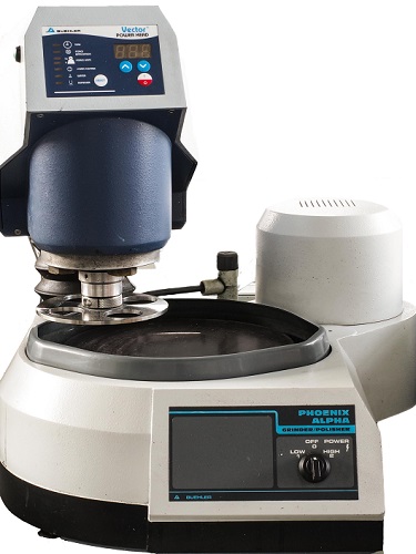
Bühler Alpha Phoenix

Grinding / Polishing

The samples produced must be grinded and polished so that they can subsequently be subjected to microstructural analysis.

Technical data:
Bühler Alpha Phoenix

• Disc diameter: 250mm
• Speed: 150-300rpm
• Number of samples: max. 4
• adjustable polishing pressure
• different polishes

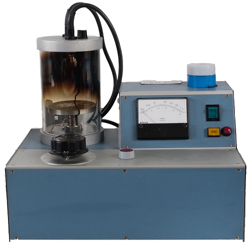
Steaming

This allows non-metallic surfaces to be made conductive by vapor deposition with carbon so that analyses can be carried out under a scanning electron microscope. Furthermore, special material analyses can be carried out.

Technical data:
Balzer Union CED 010

• Vacuum: 0.05 mbar
• Material for steaming: carbon yarn

Balzer Union CED 010
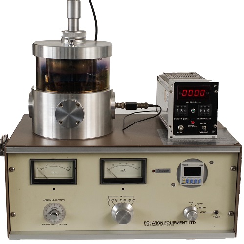
Palaron Equipment SEM Coating E5100

Sputtering

It is possible to sputter samples with gold to make them conductive for scanning electron microscopy.

Technical data:
Palaron Equipment SEM Coating E5100

• Process gas: Argon
• Medium: Gold target

Microscope

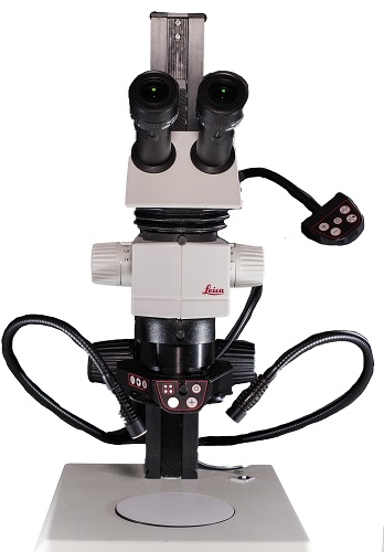
Leica M80

Stereo zoom microscope

A simple microscope to get a first overview. Preparations of the samples can be performed here using tweezers and a scalpel.

Technical data:
Leica M80

  • Magnification: max. X6
  • Illumination: LED ring light and gooseneck

System microscope

With this system microscope, bright/dark field contrast images can be acquired. This means that initial microstructure analyses can already be performed here.

Technical data:
Olympus BX 40

  • Max. enlargement: X1000
  • Max. resolution: 0.27 µ
  • Focusing accuracy: approx. 1 µ
  • Illumination: Köhler illumination for incident light
  • 5-fold objective nosepiece
Olympus BX 40
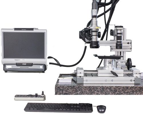
Keyence VHX-700F

Digital microscope

Due to its different lenses, the digital microscope is a universally applicable microscope, which can be used for simple visualizations up to microstructure analyses. It is possible to carry out measurements, which is essential for detailed protocolling.

Technical data:
Keyence VHX-700F

  • 1/1/8 CCD image receiver
  • Max. resolution: 2 mio. pixels
  • 3D representation of the surface
  • Close range lens: enlargement X5 – X50
  • Zoom lens: enlargement X20 – X200
  • Universal zoom lens: enlargement X100-X1000
  • High resolution zoom lens: enlargement X500-X5000

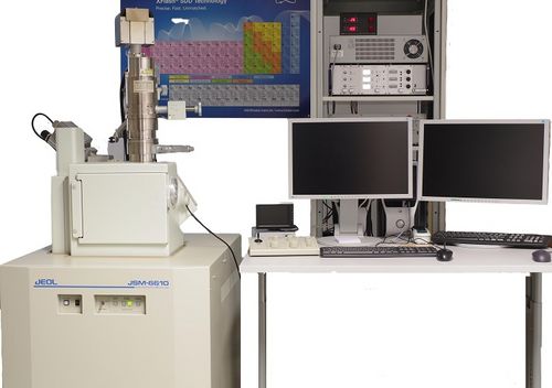
Scanning electron microscope

The scanning electron microscope is one of the high-resolution microscopes that have a high depth of sharpness despite great enlargements. With this microscope, microstructure and material analyses can be created.

Technical data:
Joel JSM 6610 SE

  • Cathodes: Tungsten, LaB6
  • Resolution: approx. 0.5 nm at 5kV
  • Enlargement: X5 – X300000
  • Detectors:
    • Secondary electron detector (SE)
    • Backscatter electron detector (BSE), for material contrast imaging
    • Energy dispersive X-ray analysis (EDX)
      • quantitative and qualitative detection of elements
      • Point and line analysis and distribution images
  • Sample table: 5-axis
  • Sample weight: max 1kg
Joel JSM 6610 SE

Prof. Dr. Stefan Trapp

Professor Department of Environmental Planning/Environmental Engineering – Physics and Power Engineering

LOCATION

Birkenfeld | Building 9916 | Room 118

Homepage

Steven Hohmann

Employee of Environmental Planning/Environmental Engineering Faculty – Department of Mechanical Engineering